Dear Doctors: My wife is 59 years old. She had COVID-19 but recovered. A recent chest X-ray shows a white patch on her lung. She doesn’t have a cough or a fever, and her pulmonologist wants her to have a PET scan. What is that? Are PET scans safe?
Dear Reader: A PET scan is a type of imaging test that creates 3D pictures of the interior of the body. The acronym stands for positron emission tomography. “Positron” refers to the subatomic particles involved in the test, and “tomography” means that the resulting images are presented as cross-sectional slices.
PET scans examine the tissues of the body at a cellular level. Because these tests can measure metabolic activity, such as oxygen use, blood flow and the rate at which specific areas of the body are using glucose, they are used to evaluate organ function and detect the onset of disease at an early stage. PET scans are commonly used to identify brain disorders, heart problems and cancers, and to evaluate the progress of cancer treatments.
The test begins with the injection of a small amount of a substance, such as glucose, oxygen, carbon or nitrogen, that is naturally used by the part of the body being examined. That substance, known as a tracer, has been tagged with a tiny amount of short-lived radioactive material that emits positrons. It takes about an hour for the tracer to circulate throughout the body, during which time the person is asked to rest quietly.
Cancer cells use glucose at a higher rate than normal cells. When glucose is used as the base of a tracer, potential tumors appear brighter than the surrounding tissues in the resulting scan. However, slow-growing tumors, which don’t use glucose as quickly, may not appear at all. Using the other types of tracers, PET scans can identify blockages in the heart and cardiac vessels and highlight abnormalities in brain structure, as occur in Alzheimer’s disease, epilepsy or Parkinson’s disease.
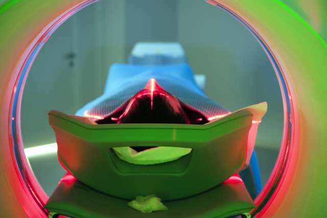
The scan itself takes place using a machine that looks a bit like a series of large doughnuts with a bed sliding through them. The person lies on the flat bed and stays as still as possible as it slowly glides through the round opening. The doughnut contains cameras that detect and record emissions from the radioactive tracers. Multiple images are produced in slices, which are then compiled into a 3D picture of the body. These are used to aid in diagnosis and guide treatment. The scan itself takes 20 to 30 minutes.
PET scans are considered to be safe. The main risk arises from exposure to radiation during the test. That exposure is small, equal to about three years’ worth of natural background radiation. The injected radioactive substances have a very brief lifespan, measured in hours, and are completely excreted via the urine and feces within a day. Drinking fluids helps speed that process. If you or your wife are concerned, your doctor can let you know if a different scanning procedure may be an option.
(Send your questions to [email protected], or write: Ask the Doctors, c/o UCLA Health Sciences Media Relations, 10960 Wilshire Blvd., Suite 1955, Los Angeles, CA, 90024. Owing to the volume of mail, personal replies cannot be provided.)