Diseases of the brain can be among the most difficult to treat, in part due to the protective nature of the blood-brain barrier. UCLA neurosurgeons are using the most advanced forms of neurotechnology to address these challenges in the clinic and in clinical trials.
Many of these advances have involved ultrasound, which uses high-frequency sound waves to produce real-time images, is safe, easy to use, and has dramatically improved in recent years, making it an attractive option for diagnosing disease and guiding medical procedures.
“Now, what we can see with ultrasound is close to the level of detail we can see with MRI (magnetic resonance imaging),” says Richard G. Everson, MD, an assistant professor of neurosurgery at the David Geffen School of Medicine, who specializes in treating tumors of the brain, spine, and skull base. “With ultrasound, you can use it in the operating room to see things change in real-time.”
UCLA Health neurosurgeons are pushing the boundaries of ultrasound to make the removal of brain tumors safer and more effective.
One of the latest advancements is navigated intraoperative ultrasonography.
Removing a brain tumor is a delicate balancing act. Determining where healthy tissue ends and cancerous tissue begins is especially challenging. Surgeons risk removing too much and cutting into healthy tissue – or taking out too little and leaving behind dangerous cancer cells.
Neurosurgeons typically rely on Magnetic Resonance Imaging (MRI) to provide the detail and resolution required to delineate the borders of a brain tumor. However, “when you open up the skull, the brain can shift, or it can swell. So, the map of the brain based on the pre-operative MRI is no longer accurate,” Dr. Everson says.
Obtaining an MRI in the middle of surgery requires the construction of specialized MRI suites, and can add several hours to a surgery, increasing patient risk.
Navigated intraoperative ultrasound combines the best of both worlds by linking the pre-operative MRI to real-time, high-resolution images from modern ultrasound units.
The images from the ultrasound get superimposed with the pre-operative MRI to allow a real-time comparison of the tumor before surgery, its shift in position during the operation, as well as the matter that has been removed and that which remains. This allows the neurosurgeon to use an ultrasound wand during surgery to more precisely match up their imaging with the operation sites.
“With our navigated ultrasound system, I can monitor the progress in removing the tumor throughout the operation, in seconds, as often as I would like, and at no added risk to the patient,” Dr. Everson says.
UCLA is one of a few centers nationally to adopt the system.
“We’ve incorporated it into our workflow for every surgery,” says Dr. Everson, “and it has enhanced accuracy, effectiveness and safety of our operations without having to make compromises.”
Visualizing brain function
Another advancement UCLA neurosurgeons are taking advantage of is functional ultrasound.
Still in its experimental stages in the context of brain surgery, functional ultrasound helps clinicians monitor brain activity. Rather than creating still images of the brain, functional ultrasound tracks minute differences in the brain’s blood flow. These can be used to visualize the brain’s hemodynamic response, in which blood flow increases to areas of the brain with high neuronal activity.
“We can look at someone’s response in very specific parts of the brain while they’re doing certain tasks, and repeat this, non-invasively, over time. That isn’t logistically feasible with MRI,” Dr. Everson says.
One clinical study at UCLA looks at patients who are recovering from surgery after traumatic brain injury and undergoing functional ultrasound over a series of weeks and months. This is an ideal population to study, since ultrasound is unable to image through the skull (patients have had part of their skull removed post-surgery).
Dr. Everson says that one day, functional ultrasound may be used to gauge recovery in patients with brain injuries or brain tumors, especially those who are unable to perform certain tasks like talking.
“Ideally, we could see the more subtle improvements in brain function that might be missed by a standard clinical exam,” he says. “We could say, ‘We see that the brain is more active here, and less active there,’ so maybe we could do something to stimulate that region to wake it up.”
Treating disease
However, ultrasound isn’t only helping surgeons to guide procedures and monitor recovery – it’s also being used to treat conditions such as essential tremor and Parkinson’s disease.
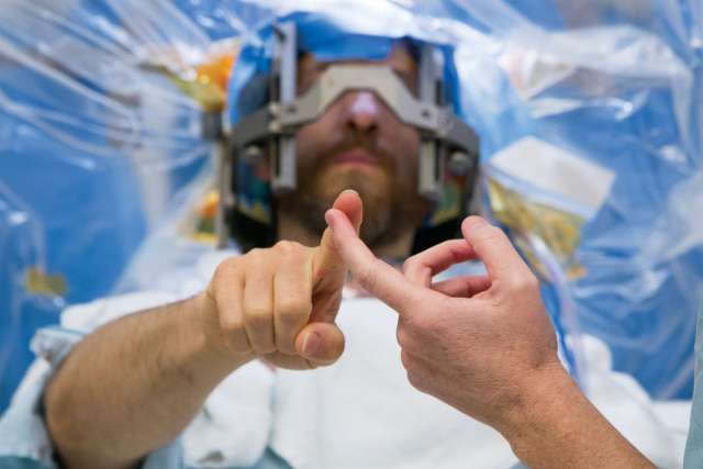
UCLA Health has been at the forefront of adopting MRI-guided focused ultrasound to treat tremor, adopting the technology soon after the U.S. Food and Drug Administration approved it in 2016.
More on MRI-guided focused ultrasound
MRI-guided focused ultrasound takes advantage of the fact that acoustic waves can be focused precisely and converted into heat. That heat can be used to burn or “lesion” a specific area of the brain involved in producing disabling tremor.
This allows the surgeon to significantly reduce or eliminate tremor without the need for a surgically placed brain implant.
UCLA Health neurosurgeons are using the technology as a non-invasive, non-surgical approach to treat tremor in patients with Parkinson’s disease, essential tremor and other diseases in patients that are not adequately treated with medications.
MRI-guided focused ultrasound is also being tested at UCLA as both a diagnostic and therapeutic tool in patients with brain cancer.
The blood-brain barrier, which protects the brain against viruses and bacteria, also prevents some cancer drugs from reaching brain tumors. With focused ultrasound, directing a mild amount of sound waves into brain tissue can temporarily and safely open the blood-brain barrier, allowing treatments to reach the tumor.
While the barrier is disrupted, proteins and molecules from the tumor can become measurable in the bloodstream, allowing surgeons to also take a liquid biopsy rather than obtaining a tissue biopsy with an invasive approach.
UCLA Health is currently enrolling patients with certain forms of brain cancer into a clinical trial to test the safety and effectiveness of focused ultrasound.
“Focused ultrasound could conceivably replace having to do an invasive biopsy or be included in the standard treatment of brain tumors,” Dr. Everson says.
Brain stimulation to reduce back pain
While many people think of back pain as the result of injury to muscles or joints, a growing amount of scientific evidence suggests that chronic pain can be treated by targeting an unexpected location: the brain itself.
“Pain is an emotion, and emotions come from the brain,” says Ausaf Bari, MD, PhD, a neurosurgeon at UCLA Health. “We've started to understand that in people who have chronic pain, the brain has been altered in ways that make it difficult to treat and this likely to due to changes in the emotional circuits.”
In 2020, lower back pain affected more than 600 million people globally. Many patients with chronic back pain end up turning to long-term opioid use with little success when treatments like surgery or physical therapy fail to cure their symptoms.
Deep brain stimulation (DBS) is a treatment that has been approved by the U.S. Food and Drug Administration since the 1990s to treat movement disorders, such as Parkinson’s disease, as well as to reduce seizures in patients with epilepsy.
To deliver the treatment, neurosurgeons implant electrodes in certain areas of the brain, which produce electrical impulses that can correct abnormal rhythms or circuitry. The amount of stimulation that is delivered is controlled by a device that works like a pacemaker, which is placed under the skin in a patient’s chest.
Now, with funding from the National Institutes of Health’s Helping to End Addiction Long-term (HEAL) initiative, Dr. Bari is leading a study to determine whether DBS could help cure lower back pain that has failed traditional surgical management.
In a proof-of-concept pilot study, Dr. Bari and his team are aiming to demonstrate the safety and feasibility of DBS for chronic low back pain, as well as to better understand the potential mechanisms of the treatment and identify neuroimaging biomarkers to measure its effectiveness.
“So far, we’ve safely implanted the device into one patient, and he’s been very happy with the results,” Dr. Bari says. “Ultimately, I don’t think DBS is going to be the only solution to back pain, but it could be part of a comprehensive treatment plan potentially allowing other therapies like physical therapy to work better.”