Aortic Care
Our experts have decades of experience providing advanced treatments for aortic disease. We perform all types of endovascular and aortic valve repair procedures.
Why choose UCLA Health for aortic care?
The UCLA Health Aortic Center is a recognized Center of Excellence in aortic care. Patients from all over the world come to us for our expertise. Our specialists have decades of experience treating aortic disease. We don’t just use the latest techniques – in many cases, we helped develop them.
Additional highlights of the Aortic Center include:
Treatment from leading experts: Specialists in the UCLA Health Aortic Center have made some of the most significant breakthroughs in treating aortic disease. For example, we helped develop a surgery called hybrid repair. And we performed some of the first minimally invasive repairs in the country. We are also one of only a few centers in the U.S. that offers a highly specialized minimally invasive surgery for bulges in the bend of the aorta (aortic arch aneurysms).
Patient-centered, convenient care: Our specialists create personalized treatment plans. We focus on lifestyle changes as well as medical management for a comprehensive care approach. With on-site advanced imaging, we can coordinate your full medical evaluation efficiently, often in a single office visit.
Team approach: Our care team includes specialists in cardiac surgery, vascular surgery, anesthesiology, cardiology and radiology. This coordinated approach allows us to offer comprehensive diagnosis and a well-rounded treatment plan.
Minimally invasive techniques: We can use a minimally invasive approach for most aortic problems, meaning fewer patients need open surgery. These treatments also mean less pain, faster recovery and a shorter hospital stay.
Aortic conditions we treat
The aorta is our largest artery, responsible for carrying oxygen-rich blood throughout the body. Conditions that affect the aorta can lead to life-threatening complications. The UCLA Health Aortic Center is world-renowned for the diagnosis, evaluation and treatment of all types of aortic diseases.
In particular, we specialize in the treatment of:
Aortic aneurysms: Irregular bulges in the aorta, which can split the artery walls (dissection) or burst (rupture)
Aortic dissections: Tears in the aorta’s walls, leading to blood leaks that can be life-threatening
Aortic occlusive disease: Blocked blood flow in the aorta
Aortic valve disease: A condition in which the valve between the heart’s pumping chamber (left ventricle) and the aorta doesn’t work correctly
- Aortic regurgitation: When the aortic valve doesn’t close properly, causing blood to leak backward from the aorta to the left ventricle
- Aortic stenosis: When the aortic valve narrows, restricting proper blood flow
Traumatic aortic injuries: Injuries that cause a puncture, tear or bruise on the aorta, often occurring because of a motor vehicle accident or gunshot wound
Additional conditions we treat include:
Aortic arch aneurysms: Irregular bulges in the bend of the aorta (aortic arch)
Aortitis: Inflammation in the aorta
Bicuspid aortic valve: An aortic valve that only has two flaps instead of the usual three, often leading to valve narrowing (stenosis)
Marfan syndrome: A genetic disorder that affects connective tissue, often leading to an aortic aneurysm
Tests & procedures we offer
Our specialists offer a range of services to diagnose and treat aortic conditions, including:
Advanced imaging
Accurate diagnosis starts with advanced imaging. At the UCLA Health Aortic Center, we use state-of-the-art technology that offers unmatched precision in detecting aortic disease. Imaging options include:
Angiography: The doctor uses a small flexible tube (catheter) to send a contrast dye to the coronary arteries. With X-rays, the doctor can track the flow of the contrast dye through the arteries and aorta to detect blockages.
CT scan: These tests use computers and low doses of radiation to capture reliable images of the aorta. We can also combine CT scans with contrast dye to get higher-resolution images of the aorta.
Dual-source CT scan: We were among the first centers in the country with an imaging machine called a dual-source 128-slice CT scan. This machine allows us to quickly capture detailed images with significantly reduced radiation doses.
Duplex ultrasound: This test combines traditional sound wave technology with a Doppler ultrasound. Using an ultrasound wand on the outside of the chest, the doctor tracks the movement and speed of blood flow through the aorta.
Intravascular ultrasound (IVUS): With IVUS, the doctor places an ultrasound device inside the artery to examine the aorta from the inside out. This allows us to view high-definition images of aortic ulcers, aneurysms or dissections.
MRI: This noninvasive scan uses powerful magnets and radio waves to obtain highly detailed images. We use advanced MRI techniques to evaluate blood flow in the aorta and branch vessels.
Nuclear perfusion imaging: This test enables doctors to compare how the heart performs at rest with how it acts under stress. The test involves injecting a safe radioactive tracer into the patient’s bloodstream, then using imaging to view how and where the tracer travels.
Minimally invasive treatment for aortic disease
Our specialists offer a full range of minimally invasive treatments, many of which were developed in our operating rooms. We perform:
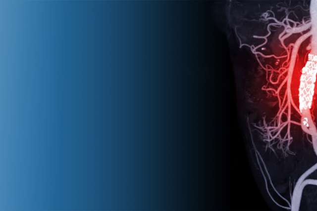
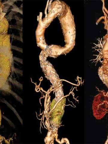
Endovascular aortic repair (EVAR): Surgeons use EVAR to treat an abdominal aortic aneurysm by placing a small mesh tube (stent graft) in the damaged part of the aorta. The surgeon places the stent using a thin tube (catheter) inserted into an artery through small incisions in the groin.
Fenestrated endovascular aortic repair (FEVAR): Surgeons use FEVAR when an aneurysm or dissection occurs where the blood vessels from the abdominal aorta (renal arteries) branch off to the kidneys. Fenestrated stent grafts have holes that line up with these arteries. The holes allow blood to flow to the kidneys and other organs.
Hybrid repair: This two-stage procedure combines a less-invasive open surgery with an endovascular approach. The surgeon uses open surgery to restore blood flow, and then uses a minimally invasive approach to repair the aorta. Doctors perform this surgery on patients who are too high-risk for a traditional open procedure.
Transcatheter aortic valve replacement (TAVR): TAVR replaces an aortic valve with severe stenosis. The surgeon inserts a thin tube (catheter) into an artery in the groin and implants the valve through the catheter.
Meet our team
Our vascular surgeons and cardiothoracic surgeons are leaders in aortic disease treatment. We’re renowned for our expertise in performing the latest minimally invasive endovascular techniques. In many cases, we even helped develop them.
Aortic Physicians at UCLA

Contact us
Call to request an appointment with an aortic specialist at UCLA Health.
Find your care
Our specialists are experts in treating aortic disease. To learn more about our services, call .
